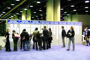
| <Certifificate of Merit> |
“ What Happen?”CT Imaging Spectrum after Radiofrequency Ablation of Lung Malignancy : Typical and Atypical Findings
郷原英夫氏(岡山大学医学部・歯学部附属病院放射線科) |
| |
Emergency CT Examination: Important Issues for the Diagnosis of Gastrointestinal Perforation
荒木哲朗氏(近畿大学医学部放射線医学教室診断学部門) |
| |
Dual Source, Dual Energy CT Imaging of the Liver : Current Status and Further Prospects
北野 悟氏(奈良県立医科大学放射線科) |
| |
How to Use Liver-Specific Contrast Agents on MRI and Sonography : Enhancement Patterns of Kupffer, Sinusoid, and Hepatocyte Using Superparamagnetic Iron Oxide, Gd-EOB-DTPA, Galactose-Palmitic Acid and NC100100
岡田真広氏(近畿大学高度先端総合医療センターPET 診断部門) |
| |
Clinical Utility of 3T Dynamic 3D MRI with MPR, CPR, MIP, and Volume Rendering : Technique for Liver, Biliary, and Pancreatic Disease
久能由記子氏(久留米大学医学部放射線科) |
| |
Real-time CAD System for Automated Detection of Calcifications in Breast Ultrasonography
石原福太郎氏(岐阜大学)ほか |
| |
Clinical Value of 3D Fused Image of Myocardial Perfusion SPECT and CT Coronary Angiography
田代城主氏(熊本大学大学院医学薬学研究部放射線診断学) |
| |
Malignant Lymphoma Post Therapy : Usefulness and Pitfalls Using FDG-PET/CT Compared to CT, MRI and Ga Schintigraphy
岡田真広氏(近畿大学高度先端総合医療センターPET 診断部門) |
| |
Visualization of Endolymphatic Hydrops in the Patients with Meniere’s Disease by MR Imaging after Intratympanic Administration of Gd-DTPA : Its Methods, Anatomy, Findings, and Clinical Application
長縄慎二氏(名古屋大学大学院医学系研究科量子医学分野) |
| |
Diversity in“Axial Plane”for Brain MRI
田岡俊昭氏(奈良県立医科大学放射線医学教室) |
| |
MR Imaging of Major Salivary Gland Masses and Pathological Correlations
本折 健氏(千葉大学医学部附属病院放射線科)ほか |
| |
Head and Neck MR Imaging with Advanced Techniques
藤田晃史氏(自治医科大学放射線医学教室) |
| |
Paranasal Sinus Imaging; Virtual Visualization for Endoscopic Sinus Surgery(ESS)
鈴木晶子氏(横浜市立大学医学部放射線科) |
| |
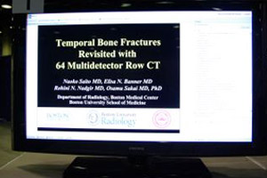
Sickle Cell Disease : Radiographic Manifestations in Head and Neck
斎藤尚子氏(Department of Radiology, Boston Medical Center, Boston University School of Medicine/埼玉医科大学放射線科)ほか |
| |
Important Anastomotic Pathways between External Carotid Artery(ECA)and Intracranial Arteries
村田隆紀氏(東北大学医学部放射線診断科) |
| |
Uterine Pathologies in 3T-MRI : Clinical Application of Diffusion-weighted Imaging and MR Spectroscopy in Differentiating Benign and Malignant Lesions
竹内麻由美氏(徳島大学医学部放射線科) |
| |
Susceptibility-weighted Magnetic Resonance Imaging : Assessment for Intracranial Venous System and Hemorrhagic Components in Neonates and Infants
丹羽 徹氏(神奈川県立こども医療センター放射線科) |
| |
Radiation Protection Basics for IVR Staff : Usefulness of Non-lead Aprons
千田浩一氏(東北大学大学院医学系研究科保健学専攻) |
| |
Angiographic Findings of Anatomical Changes of Hepatic Artery after TACE in Patients with HCC
末吉英純氏(長崎大学医学部歯学部附属病院放射線科) |